Advances in Neutrophil Testing In Type 2 Diabetes Mellitus-Juniper Publishers

Authored by Bernhard Otto Boehm
Abstract
Patients with type 2 diabetes mellitus (T2DM) suffer
from impaired glucose metabolism which results in low-grade inflammation
and activation of the innate immune system. Neutrophils the key
effector cells of the innate immune system and heavily implicated in the
pathogenesis of T2DM, are promising cell-based inflammatory biomarkers
for immune health profiling, provided that they can be rapidly purified
and measured with sufficient precision. In this review, we highlight
recent advances in neutrophil isolation and functional assay using
microfluidics technologies and the potential of their functional
phenotype as a novel biomarker of vascular risk in diabetes.
Keywords: Neutrophils; Diabetes; Point-of-care; Microfluidics; Immunology
Introduction
With the increasing aging population worldwide,
metabolic disorders such as diabetes mellitus (DM) and cardiovascular
diseases (CVDs) have become the main public health challenges with
rising premature morbidity and associated mortality, as well as
escalating healthcare costs [1].
DM is characterized by chronic hyperglycemia resulting in increased
oxidative stress, inflammation and endothelial dysfunction [2,3].
Patients with CVDs or type 2 diabetes mellitus (T2DM) often exhibit
low- grade inflammation, and are assessed based on established
cardiovascular risk factors (glycemic control, blood pressure and
lipids). Immune health is evaluated by differential leukocyte count and
circulating biomarkers (cytokines and C-reactive protein (CRP), which
are suboptimal for monitoring stage- dependent pathogenesis, advocating
the need to develop new cell-based biomarkers that can quantity specific
immune functions in addition to leukocyte enumeration.
Neutrophils, the key effector cells of the innate immune system, play a pivotal role in T2DM and CVDs pathogenesis [4]. Various neutrophil dysfunctions have been reported in T2DM patients including cell stiffening [5,6] impaired chemotaxis [7,8] and phagocytosis which lead to increased susceptibility to bacterial infections [9].
Despite the adverse changes of leukocytes in diabetes, there are
currently no specific measurements to assess patient's leukocyte
phenotypes or inflammatory status. As distinct neutrophil subsets
exhibit functional and phenotypic differences [10]
a better understanding of their phenotype and pathophysiological
relevance requires novel neutrophil separation tools (independent of
surface markers) to improve their predictive capabilities as novel
biomarkers [11].
Microfluidics, also known as "lab-on-a-chip" technologies, is a
powerful toolbox for rapid sample preparation and detection with its low
consumption of sample and reagents, device miniaturization, and
single-cell analysis [12].
In this short review, we will highlight recent advances in
microfluidics- based neutrophil testing technologies, and the potential
of neutrophilfunctional phenotype as biomarkers for diabetes testing.
Discussion
Neutrophil isolation
Neutrophil polymorphonuclear granulocytes (PMN) are
the most abundant leukocytes (~50-70%) in humans, with ~2-5x10⁶
neutrophils per mL of whole blood (~109 RBCs). They are short-lived
(~5-24hr), prone to activation [13]
and should be processed quickly within 2-4 hours of collection.
Conventional neutrophil isolation methods include density gradient
centrifugation and RBCs lysis, which are laborious (~1- 3hr) and require
large blood volume (~10mL). Commercial kits based on magnetic
bead-based affinity binding (MACS xpress® Neutrophil Isolation Kit
(Miltenyi Biotec) and Easy SepTM neutrophil enrichment kit (STEMCELL
Technologies) provide high neutrophil yield and purity by negative
selection, but is expensive for large volume processing.
Microfluidics technologies for neutrophil isolation
have been developed based on affinity binding to functionalized surfaces
using common neutrophil markers (CD66b, P-selectin) [14,15].
However, these methods require on-chip cell analysis as it is
non-trivial to elute the purified neutrophils off-chip for downstream
assays. Our group has previously developed an efficient size-based cell
sorting technique known as Dean Flow Fractionation (DFF) based
oninertial focusing phenomenon in micro channels [16].
In DFF systems, fluid flowing through a curvilinear (spiral) channel
experiences centrifugal acceleration directed radially outward, leading
to the formation of two counter-rotating vortices known as Dean vortices
[17].
Besides inertial lift forces (FL) particles experience lateral Dean
drag force (FD) due to these transverse Dean flows, which results in
superior separation resolution as both forces (FL and FD) scale
non-linearly with particle size [18-20].

We first applied DFF technology to isolate circulating tumor cells (CTCs) [21]
and microorganisms from whole blood whereby the size ranges of the
target cells are distinctly different from RBCs. By exploiting the
subtle size differences between major leukocyte subtypes
(neutrophils/monocytes~10-12μm lymphocytes~7-9μm), we recently developed
a novel DFF spiral micro device to purify neutrophils rapidly from
whole blood for functional phenotyping in T2DM [22].
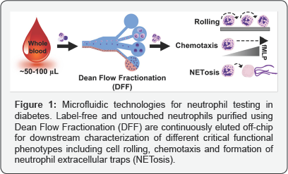
The developed technology enables single-step neutrophil isolation
(>90% purity) without immune-labeling, saving both time and cost. In
addition, the sorted "untouched" neutrophils are continuously eluted
off-chip with simultaneous buffer exchange, facilitating user operation
and eliminating the need for centrifugation. Moreover, as the method
only requires small blood volumes (finger prick ~50- 100μL) it can be
easily integrated with other cellular assays or detection modules for
point-of-care (POC) testing (Figure 1).
Neutrophil rolling
During endothelial inflammation, leukocyte adhesion
cascade is a multi-step process involving cell rolling, adhesion and
transmigration through blood vessel walls to the site of injury [23].
Neutrophil rolling is widely considered a critical step as it can
affect cell adhesion with impaired cell tethering or increased rolling
speeds [24,25].
Several microfluidics- based cell rolling assays have been reported
previously to study rolling behavior under physiological flow conditions
(~1-10dynecm-2), but not in disease-specific context [26-28].
In our study, we combined DFF neutrophil sorting method and
microfluidics assay to measure neutrophil rolling speed on E-selectin, a
cell adhesion molecule expressed by activated endothelium to initiate
leukocyte recruitment. This neutrophilendothelial interaction is
mediated by several sialyl Lewisx presenting ligands expressed on
leukocytes including P-selectin glycoprotein ligand 1 (PSGL1),
glycosylated CD44 and E-selectin ligand 1 (ESL1) [29].
In our clinical validation, we observed a significant
down regulation of neutrophil PSGL-1 expression in T2DM patients. Using
automated cell tracking algorithm, we further showed that rolling
trajectories ofT2DM neutrophils were more discontinuous and irregular as
compared to healthy neutrophils. Interestingly, diabetic neutrophils
had ~20% higher rolling speeds, which correlated with neutrophil
activation, PSGL-1 expression, as well as established cardiovascular
risk factors (cholesterol, CRP and HbA1c). Taken together, the data
support the hypothesis that neutrophil-endothelial interactions are
impaired in T2DM patients which can lead to defective neutrophil
recruitment, and thus increased patient susceptibility to infection.
Neutrophil chemotaxis
Chemotaxis, a dynamic process where cells sense and
move in response to chemical gradients, is traditionally studied using
Boyden chamber (transwell), Dunn chamber and micropipette assay [30].
However, these methods suffer from poor reproducibility and ill-defined
chemical gradients, which could be overcome by using microfluidics
technologies to generate stable and linear chemo attractant gradient in
small length scale (~μm) [15].
Moreover, most microfluidic chemotaxis assays only require ~102-3
neutrophils, and facilitate real-time imaging of cell movement at single
cell resolution [31].
First performed clinical testing of patients with burn injury using
microfluidics, and observed that neutrophils suffered from impaired
directionality or slower migration speed, which were associated with
degree of burn injury.
Similarly neutrophils from asthmatic patients also
displayed significantly slower migration speed as compared to healthy
subjects, suggesting its use as a novel diagnostics marker [32].
As impaired neutrophil chemotaxis behavior was reported previously in
diabetic patients our group has developed an integrated micro device for
neutrophil chemotaxis assay using a drop of blood. The novelty lies in
the single-step enrichment of neutrophils using biomimetic cell
margination [33]
and affinity capture, followed by simultaneous exposure to chemotactic
gradient without requiring additional user manipulation [34].
In our preliminary clinical data we also observed signification
suppression of chemotaxis behavior in T2DM patient, which can be
mitigated by short exposure to metformin in vitro. Besides diagnostics
applications, microfluidics chemotaxis assays also enable study of
complex chemoattractant gradients with high precision [35], well-controlled spatial and temporal gradients to probe cell migration pattern [36,37], as well as effect of inflammatory mediators in neutrophil-monocyte interactions [38].
Neutrophil extracellular traps (NETs)
First discovered in 2004, formation of neutrophil
extracellular traps (NETs) is an innate key defense mechanism against
bacterial infections through the release of nuclear and granular
contents to contain and kill pathogens [39].
Upon activation or exposure to bacteria, histones undergo
citrullination, followed by chromatin decondensation. Nuclear membrane
will degrade, leading to DNA release into the cell, and subsequently
extrusion out of neutrophils. Secreted NETs (process known as NETosis)
then form a sticky scaffold consisting mainly of microbicidal
proteases/elastase and cytotoxic molecules (histones). Interestingly,
recent work have shown that diabetic neutrophils were more susceptible
to NETosis [40], which can mediate delayed wound healing [41].
NETs components (elastase, histones, neutrophil
gelatinase- associated lipocalin, and proteinase-3) are also elevated in
the blood of patients with diabetic foot ulcers, and were associated
with infection or worsening of ulcer [42].
Overall these clinical evidences suggest a major role of NETosis in
diabetes pathophysiology and endothelial damage making it a novel
biomarker for early detection of diabetes-related vascular or end-organ
complications. Compared to chemotaxis development of microfluidics NET
osis assay is still at its early infancy with a recent reported assay
based on fluorescent imaging of nucleus degradation [43].
Nevertheless given the increasing importance of NETosis and easy
quantification using imaging, we expect more development of novel tools
to measure NETosis phenotype in POC settings.
Conclusion
Multidimensional neutrophil phenotypic markers will
significantly improvetheir predictive capabilitiesasinflammatory
biomarkers provided that they can be rapidly purified and measured with
sufficient precision. Microfluidics technologies are not only useful for
efficient neutrophil purification but they can also be readily
developed and integrated into POC testing plat forms to look at the sum
effects of diabetes, hypertension and hyperlipidemia. This enables
proper identification of high risk patients with appropriate follow up,
reduces the risks in different aspects of the endothelial activation
pathway and in time, the effects of therapeutics can also be studied in
diabetes and other dysmetabolic diseases.
To Know More About Current Research in Diabetes & Obesity
Journal Please click on:
https://juniperpublishers.com/crdoj/index.php
https://juniperpublishers.com/crdoj/index.php


Comments
Post a Comment